Wisconsin Equine Clinic & Hospital (WECH)
Diagnostic imaging has become an increasingly important part of veterinary medicine. Wisconsin Equine Clinic and Hospital realizes the importance of having these services available to provide the best and most efficient care to our patients. For this reason, many of these services are now mobile and can be provided at either the farm or hospital setting. MRI, for example, allows imaging of both soft tissues and bone in a way that no other diagnostic procedure can leading to a more accurate diagnosis, treatment plan and outcome.

Standing CT
Now Offering
We're excited to share with our community that Wisconsin Equine Clinic & Hospital has partnered with Asto CT to introduce cutting-edge Computed Tomography (CT) technology to our practice. Construction completed in August 2024 and we are now booking out for your horse's CT needs. WECH strives to offer compassionate care and individualized treatment to our community; with our new standing CT clients will receive a fast diagnosis through high quality images that can be acquired within 15 minutes.
Standing MRI
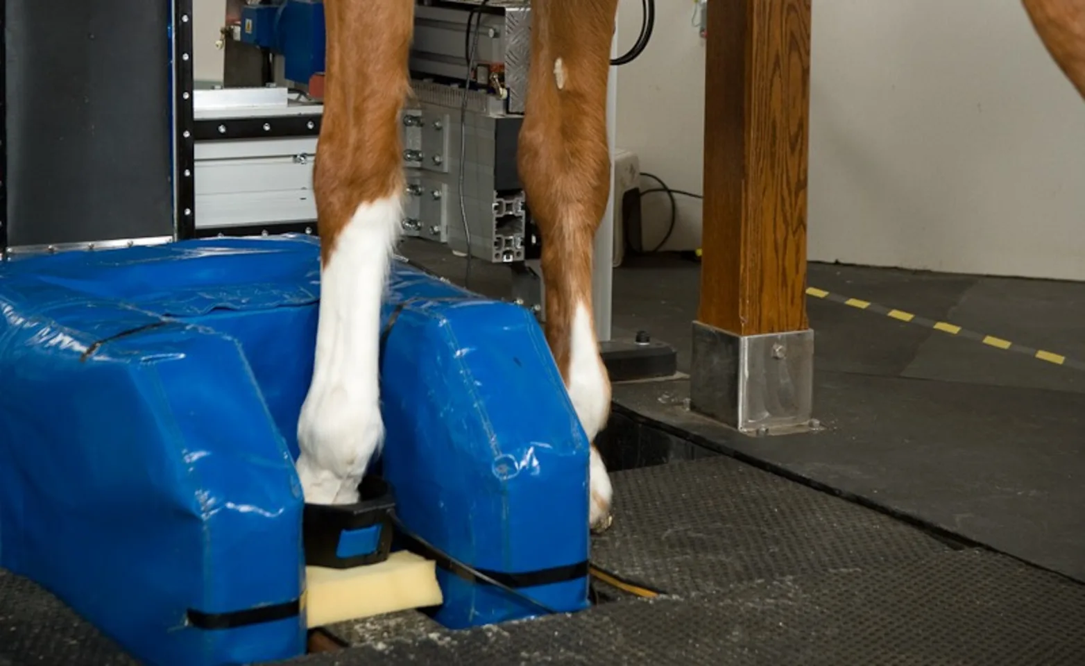
The Hallmarq Standing Equine MRI machine is specifically designed for equine patients and provides high quality images of both bone and soft tissue. During the procedure the patient stands comfortably under sedation which reduces the risks associated with general anesthesia. MRI helps to guide an accurate, early diagnosis as well as precise treatment which can decrease long term lay-up post injury.
Digital Radiography
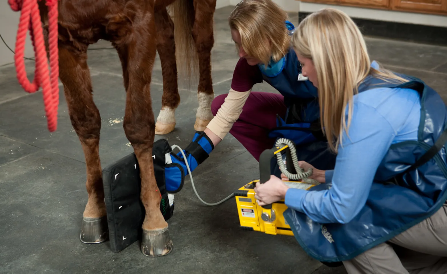
Digital radiography involves taking X-rays of the horse using specialized equipment. Unlike traditional film-based X-rays, digital radiography produces high-quality images that can be viewed instantly on a computer screen. The advantages of digital X-rays include improved image quality, efficiency, and the ability to manipulate and enhance images for better visualization. Immediate availability and the ability to enhance images is extremely important for pre-purchase exams and in treatment decisions for lameness. Applications of digital radiography in the hospital not only include musculoskeletal examination but also assessing the cervical spine, back and pelvis, the thorax for issues of the heart and lungs and abdominal x-rays looking for sand or enteroliths.
Digital Ultrasound
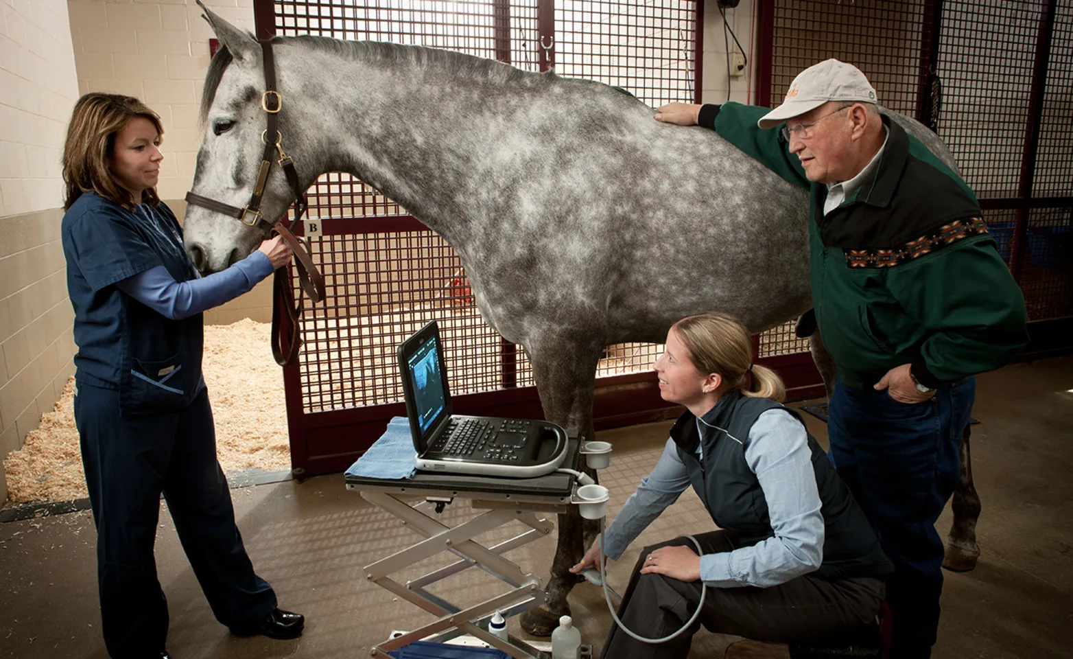
Digital Ultrasound - we offer state-of-the-art digital ultrasound services for our equine patients. Using high-frequency sound waves, we create real-time images of soft tissues allowing precise evaluation of areas such as tendons, ligaments, and internal organs. Our experienced veterinarians use digital ultrasound for lameness evaluations, reproductive health monitoring, abdominal imaging, ophthalmic exam, lung and cardiac assessment.

Endoscopy
Our team of diversely-trained equine veterinarians utilize cutting-edge technology to visualize and diagnose various conditions. We are proud to be one of the few veterinary hospitals in the Midwest to offer dynamic endoscopy, allowing us to assess airway movement and function while your horse is exercising. In addition to visualization of the airway, endoscopy allows veterinarians to visualize and diagnose conditions of the bladder, guttural pouches, GI tract, and more. Whether you’re seeking routine evaluations or addressing specific concerns, our experienced veterinarians are here to provide compassionate care for your equine.
Gastroscopy
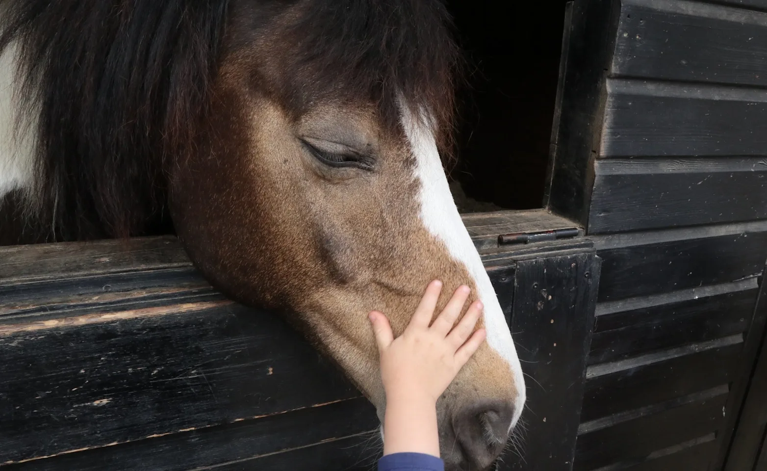
Gastroscopy is the procedure used to view the inside of a horse’s stomach. It involves passing a 10-foot long endoscope (camera) through the horse’s nose and into the stomach. This method is essential for accurately diagnosing stomach ulcers in horses, which are common and can cause signs like colic, weight loss, and poor performance. Gastroscopy can also identify tumors, stomach impactions, and other upper GI tract abnormalities as well as assist in relieving esophageal obstructions (choke). The procedure is not painful for the horse, and light sedation is used.
